“If the water is brown, turn around,” as the saying goes. Not exactly what Sera and I wanted to hear while visiting Kaua’i before I started Emergency Medicine Residency. Known as the “Garden Isle”, Kaua’i, the oldest island in the Hawaiian archipelago, is known for its lush, tropical, mountainous landscape and beaches. However, despite being a popular travel destination, there are particular dangers travelers and clinicians should be aware of.
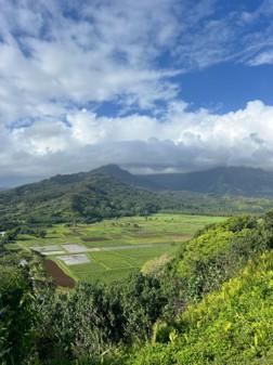
Irrigated taro fields are a source of Leptospira contamination (Photo credit: Kyle Kenyon)
Leptospira interrogans is a spirochete bacterium that can cause leptospirosis, an influenza-like illness, and in severe cases, Weil’s disease. For adventurous travelers, all is not lost. While exercising some additional precautions and remaining an informed traveler, Kaua’i has much to offer.
Leptospira and the resulting leptospirosis or Weil’s disease is a globally relevant zoonosis that can be traced back millennia. Ancient Chinese texts describe “rice field jaundice”, and in Japan, accounts of “autumn fever” can now likely be attributed to leptospirosis. In modern history, it was first recognized by Adolph Weil in 1886 while describing an unusual presentation of splenomegaly, renal dysfunction, conjunctivitis, and skin rashes – later to become known as Weil’s disease.
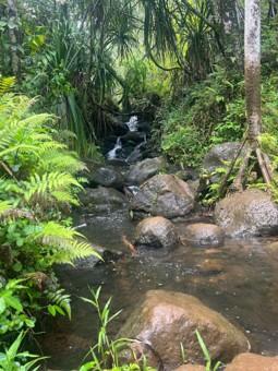
Innocuous appearing stream may contain high levels of Leptospira spirochetes (Photo credit: Kyle Kenyon)
Today, leptospirosis is one of the leading zoonotic causes of morbidity and mortality worldwide, most commonly affecting rural agricultural workers, underserved urban communities, and outdoor recreationalists. Recreational exposures include all freshwater sports including kayaking, canoeing, caving, and triathlons.
Leptospira thrives in wet tropical environments, making Kaua’i an excellent incubator. Hawaii has the highest incidence of leptospirosis in the United States – the last random fact I wanted to think about while visiting. There are features of Hawaii and Kaua’i specifically that facilitate this. Kaua’i is one of the wettest places on earth; Mount Wai’ale’ale, a shield volcano and second highest point on the island, has averaged 373 inches of rain per year over the last 30 years. After prolonged heavy rainfall, the water washes down the mountains picking up Leptospira (and other pathogens) from the soil. This is what causes the classic brown streams and ocean water that Sera and I were advised to avoid.
Animals, particularly small mammals, serve as the primary reservoir for Leptospira. Once an animal is infected, Leptospira migrates hematogenously to the renal tubules where it colonizes and is then shed in the urine. It can then survive for weeks to months in soil. Humans, technically an “accidental host”, may become infected when exposed through small abrasions or mucosal surfaces. Exposures are either direct contact with an infected animal or exposure to water contaminated with infected urine.
Once Leptospira enters the human body, it enters the bloodstream and disseminates. Leptospirosis is often described as a biphasic illness, with a brief period of apparent improvement; however, clinically these phases often blend together. Incubation occurs from initial exposure to onset of symptoms, ranging from 7 to 12 days. Initial symptoms during the first “leptospiremic phase” include fever, headache, and chills. This rather nonspecific presentation leads to missed or misdiagnoses (i.e. influenza, malaria, dengue fever). However, there are a few rather unique signs and symptoms that aid in diagnosis: muscle tenderness, particularly in the calves and lower back, and conjunctival suffusion, which is uncommon in other infectious diseases. Less common symptoms include cough and gastrointestinal symptoms. During the second “immune phase”, IgM antibodies appear in the blood and the organisms begin to deposit at their final destination. In severe cases, high levels of leptospiremia result in a sepsis-like syndrome and end organ dysfunction or failure. Severe cases are often characterized by a “cytokine storm” with high levels of various interleukins. Leptospira has a predilection for deposition in the liver, kidneys, lungs, and brain. Weil’s disease is characterized by jaundice and renal failure. Severe leptospirosis is also associated with acute respiratory distress syndrome (ARDS) and massive pulmonary hemorrhage. If not immediately recognized and treated, it can be deadly.
Diagnosis of leptospirosis is often clinical. Lab findings in leptospirosis are nonspecific but can help identify end-organ involvement. The CBC may show leukocytosis with left shift, thrombocytopenia, and anemia. Electrolyte abnormalities such as hyponatremia, hypokalemia, and hypomagnesemia are common. Renal involvement is reflected by elevated creatinine and BUN, and urinalysis may show proteinuria, pyuria, microscopic hematuria, and casts. Liver involvement shows up as elevated bilirubin, AST, ALT, and ALP. ESR, CRP, and CPK are also typically elevated.
For confirmatory diagnosis, two main pathways exist. Direct detection via PCR or dark-field microscopy can be performed on blood, urine, or CSF. Serology, including the microscopic agglutination test (MAT), is the gold-standard and works by incubating patient serum with Leptospira serovars to detect positive titers, but it can only be performed at specialized labs like the CDC and cannot be used for early diagnostic or treatment decisions.
In most cases, leptospirosis is mild and self-limiting. In an outpatient setting, patients can be prescribed oral doxycycline, azithromycin, ampicillin, or amoxicillin. Early identification and initiation of antibiotics is crucial to reduce the risk of progression to severe disease. In patients requiring hospitalization, treatment includes IV penicillin or ceftriaxone, as well as supportive measures. Leptospirosis is often associated with a potassium-wasting high-output renal dysfunction making adequate fluid and electrolyte replacement critical. In the case of complete renal failure, patients may require dialysis. Mechanical ventilatory support may be required in the case of pulmonary complications.
So, what steps can you take to protect yourself while in Kaua'i? Pay attention to local weather. The Hawaii State Department of Health will often post “brown water advisories” after heavy persistent rainfall – if the water is brown, turn around (while leptospirosis is a primary concern, brown water advisories reflect a broader contamination picture including sewage overflow, agricultural runoff, and other pathogens). Avoid swimming or wading into any freshwater sources, especially if you have any open wounds. Do not drink pond or stream water without proper treatment. Immunization is typically reserved for populations working in high-risk environments. Chemoprophylaxis and post-exposure prophylaxis with doxycycline has been shown to be effective for unavoidable exposures. Because of the high potential for exposure, all dogs should be vaccinated for leptospirosis.
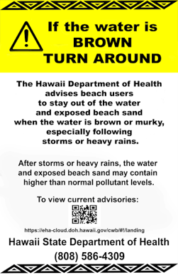
Brown Water Advisory posted by the Hawaii State Department of Health
Sera and I traveled to Kaua’i in May of 2026, towards the end of the rainy season, which typically spans from November to March. We stayed on the North Shore, which usually receives more rain than the South Shore due to the topography of the island; this is visually evident by the lushness of the north versus south. Despite being out of the rainy season, we experienced rain almost every day, mostly fleeting sun showers. Towards the end of our one week trip, there was a day of persistent rainfall. Unfortunately, our plans to surf in Hanalei Bay were canceled and I developed “Brown Water Fixation Syndrome (BWFS).” BWFS is a self-diagnosed (and made up) obsession with checking local weather advisories to try and find swimmable water. After progressing through the acceptance process, we were able to still enjoy our time in Kaua’i.
As an adventure seeker and developing Emergency Medicine Physician, I continue to work towards striking the right balance of caution and adventure. There can be multiple simultaneous truths: leptospirosis can be a serious fatal illness, most cases are mild, certain activities that may be highlights of your travel put you at higher risk, and there are steps you can take to reduce your risk. The purpose of this article isn’t to tell you what your risk tolerance ought to be, it is to give you the necessary information to make informed decisions.
Disclosures:
This article was written by the author. AI (Claude, Anthropic) was used during the editorial process to provide structural and stylistic feedback, flag clinical gaps, and answer specific questions about formatting and submission guidelines. AI was also used to summarize laboratory findings from a screenshot of a reference source, which the author then incorporated and edited into the article. All other content, ideas, clinical knowledge, personal narrative, and final editorial decisions are the author's own.